
GLAMi nomination: Be Microscopic!
institution: Corning Museum of Glass
category: Exhibition Media or Experience
https://www.youtube.com/watch?v=_vs8aV0kH8g
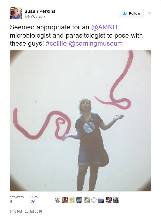
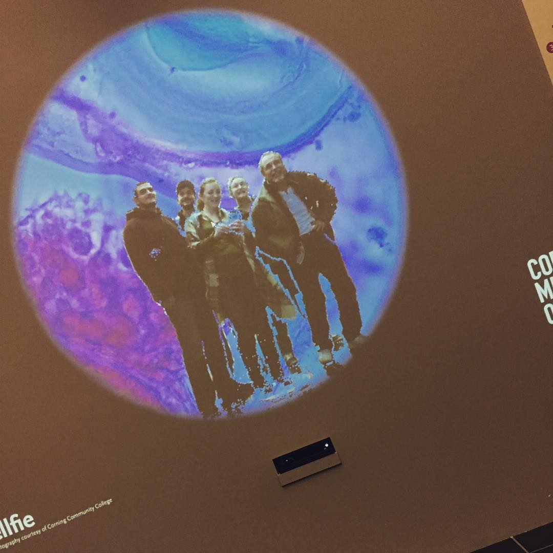
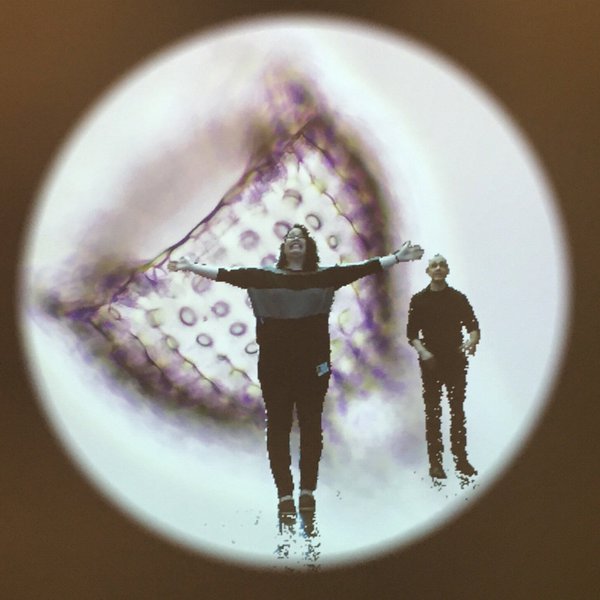
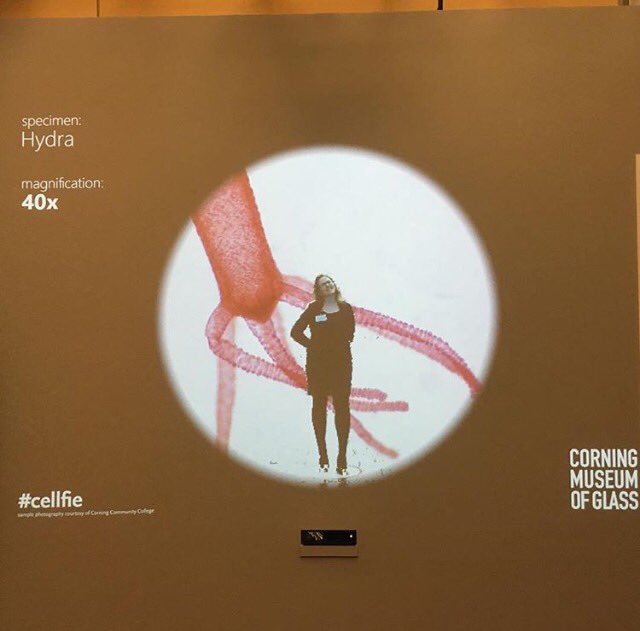
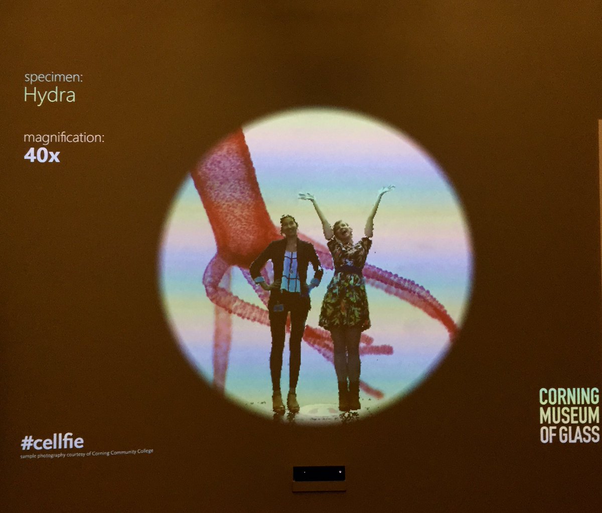
Be Microscopic! is a gestural interactive installation where users are seemingly placed under the microscope and shrunk to the size of a cell. It is installed within the exhibition Revealing the Invisible: The History of Glass and the Microscope at The Corning Museum of Glass (CMoG) on view April 23, 2016 through March 19, 2017.
Through hand gestures, users can change the specimen on the microscope slide, zoom to different levels of magnification, and initiate a freeze frame to take a #cellfie.

The interactive was developed by the museum’s digital media department using a Microsoft Kinect sensor for Windows. The device’s camera captures live video, and sensors track users’ spatial positions, body movements, and gestures. The team used a software library for Kinect called Vitruvius. This C#-based library maps the camera’s skeletal tracking data to gestural events that can be used to trigger on-screen actions.
During development, a prototype of the interactive was set up to help determine the optimum placement of the sensor, projector, and other equipment, and how to best convey to users how to interact with the program. After installation, the team observed users in action, which led to the addition of a “stand here” mark on the floor to help users find the best spot for the camera to pick up their movement, and a large icon-based gesture instruction sign hung on the top right of the interactive wall to assist users, including those who are non-English speaking.
The museum partnered with Brenda Gustin, professor of biology at Corning Community College to obtain the images of microscopic specimens for the background display. The specimens were photographed using a microscope outfitted with a digital camera by Nick Jividen, a recent graduate of the college.
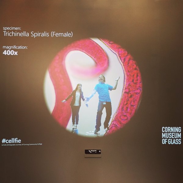
Users can shrink themselves into slides showing a Honey Bee wing, Honey Bee leg, Trichinella spiralis (female), Obelia, and Hydra. Also Radiolaria—which is significant to the museum because these microorganisms have skeletons made of silica, the main ingredient in glass. The slides show the specimens at magnification of 40x, 100x, and 400x.
As soon as a user steps into the area recognized by the Kinect, their image is shown in the interactive and the user closest to the display wall holds control of the interactions. The camera is placed beneath the display area so that users of all heights can control the interactions, making the interactive more accessible for the multi-generational visitors to the exhibition. Swiping one hand across the body progresses to the next slide, while placing both hands in front of the body and opening them increases the magnification, and closing them decreases the magnification. Waving three times activates the freeze frame, which triggers an on-screen countdown of three seconds and then the interactive display flashes and freezes so that the user can take a photo on their mobile device. Users are encouraged to share their photo with the hashtag #cellfie.
#cellfie Gallery


About the exhibition
Glass made it possible for scientists and artists to see tiny living creatures once invisible to the human eye. Revealing the Invisible: The History of Glass and the Microscope tells the stories of scientists’ and artists’ exploration of the microscopic world between the 1600s and the late 1800s. Their discoveries fed people’s hunger to learn more about nature, increasing the popularity of microscopes and driving improvements in scientific glass. These advances culminated in the 19th century with the advent of modern scientific glassmaking and the perfection of the microscope. Unleash your sense of discovery as you explore the invisible through historic microscopes, rare books, and period illustrations.